Monday, April 25, 2011

Protein in Monkeys Makes Them Resistant to HIV

Recent research at the University of Geneva and the University of Zurich have discovered that a protein called, TRIM5 is responsible for making certain species of monkeys resistant to HIV. It has been known for a few years that this protein was able to resist HIV in monkeys, but it was unclear how this was possible.


In the past few weeks recent discoveries have been made as to how TRIM5 is capable of resisting HIV. "The protein prevents the HI virus from multiplying once it has entered the cell" (ScienceDaily). This is possible because TRIM5 is able to immediately recognize when HIV enters the body and it triggers an immune response. This response is triggered through the innate immune system which differs from the adaptive immune system because it "is already able to eliminate pathogens as soon as it comes into contact with them" (ScienceDaily).


The research done at these universities has revelead how TRIM5 prevents HIV from multiplying. When HIV enters a cell, it is arranged in a complex arrangement that TRIM5 recognizes and attaches itself to. Once TRIM5 is attached it is able to trigger certain signal molecules called "polybiquitin chains" that start an anti-viral reaction to the HIV inside the cell. After this process occurs, the cell can begin to get rid of cells with HIV by "releasing messenger substances (cytokines)" (ScienceDaily).


Although this study showed that "rhesus" monkeys, also called night monkeys, were able to resist HIV, it provides new possibilities in the prevention and treatment of HIV. TRIM5 is not a protein that is unique to monkeys, humans have this protein as well. Even though it does not appear to be as effective in resisting HIV as it does in these monkeys it brings scientists closer to finding ways to fight HIV.


References:


Sunday, April 24, 2011

Nanoparticles Target Cancer Cells


By: Abby VanFossen


Sandia National Labarotories, the University of New Mexico and the UNM Cancer Research and Treatment Center have developed a method of introducing cancer killing drugs through silica nanoparticles directly to cancerous cells. These Nanoparticles are able to store large amounts and varieties of chemicals within their honeycomb shape.


The particles are described as having a nanoporous core with a high surface area and an encapsulating lipid bi-layer (liposome). The nanoparticles and the surrounding cell-like membranes formed from liposomes together become the combination referred to as a protocell: the membrane seals in the deadly cargo and is modified with molecules (peptides) that bind specifically to receptors overexpressed on the cancer cell's surface. (Too many receptors is one signal the cell is cancererous.) The nanoparticles provide stability to the supported membrane and contain and release the therapeutic cargo within the cell. The lipids also serve as a shield that restricts toxic chemotherapy drugs from leaking from the nanoparticle until the protocell binds to and takes hold within the cancer cell. This means that few poisons leak into the system of the human host, if the protocells find no cancer cells. This cloaking mitigates toxic side effects expected from conventional chemotherapy.


This method is currently being tested on human cells in vivo (occurring or carried out in a living organism) and will shortly be tested in mouse tumors. Estimates are that this method will be 10,000 times more effective than current liposome delivery methods and may be available in as early as 5 years. This will be the first work to show targetted delivery of nanoparticles to cancers supported in part by a grant from the National Cancer Institute's Alliance for Nanotechnology in Cancer .(1)


This method provides hope for easier treatment of some cancers. As well, the specific targetting that is being attempted will reduce the side effects of cancer treating drugs in the patient. This may lead to better long term health for the cancer survivor as well as less painful treatments.


1. Carlee E. Ashley, Eric C. Carnes, Genevieve K. Phillips, David Padilla, Paul N. Durfee, Page A. Brown, Tracey N. Hanna, Juewen Liu, Brandy Phillips, Mark B. Carter, Nick J. Carroll, Xingmao Jiang, Darren R. Dunphy, Cheryl L. Willman, Dimiter N. Petsev, Deborah G. Evans, Atul N. Parikh, Bryce Chackerian, Walker Wharton, David S. Peabody, C. Jeffrey Brinker. The targeted delivery of multicomponent cargos to cancer cells by nanoporous particle-supported lipid bilayers. Nature Materials, 2011


Friday, April 22, 2011

Salmonella Utilize Multiple Modes of Infection


According to the World Health Organization, the number ofSalmonella infections is continuously rising, and the severity of infections is increasing. One of the reasons for this may be the sophisticated infection strategies the bacteria have evolved. The striking diversity of invasion strategies may allowSalmonella to infect multiple cell types and different hosts. Salmonella do not infect their hosts according to textbook model. Only a single infection mechanism has seriously been discussed in the field up till now -without understanding all the details.
Extensions of the cell membrane are filled with actin filaments. In the commonly accepted infection mechanism, Salmonellaabuses the Arp2/3 complex to enter the host cell: the bacteria activate the complex and thus initiate the formation actin filaments and development of prominent membrane extensions, so-called ruffles. These ruffles surround and enclose the bacteria so that they end up in the cell interior. Last year, the research groups headed by Theresia Stradal and Klemens Rottner discovered that Salmonella can also reach the cell interior without initiating membrane ruffles. With this, the researchers disproved a long-standing dogma.
All entry mechanisms employed by Salmonella target the so-called actin cytoskeleton of the host cell. Actin can polymerise into fine and dynamic fibrils, also called filaments, which associate into networks or fibres. These structures stabilise the cell and enable it to move, as they are constantly built up and taken down. One of the most important core elements is the Arp2/3 complex that nucleates the assembly of actin monomers into filaments.
In epithelial cells, the contractile structures are less organised but work similarly. Here, actin and myosin II form so-called stress fibres that tightly connect to the membrane. During an infection, stress fibres at the entry site can contract and pull the bacteria into the cell.


Lyme Disease

Borrelia borgdorferi spirochete causes most Lyme disease in the United States. Borrelia burgdorferi is a parasite transmitted by infected ticks from deer, mice, and other small rodents that harbor the spirochete. Often these animals have a large number of the bacterium but display no symptoms. A human infected by this bacterium has widespread problems in many organ systems. The difference is the way the spirochete is transmitted and the way the immune system reacts to this bacterium. In humans, the Lyme organism has learned to survive very well.

As early as the tick bite itself, Borrelia burgdorferi can bypass the immune system in several ways. The tick has several agents in its saliva that coat the invading spirochetes, protecting them as they enter the body through the skin. This allows the bacterium to go unrecognized as a foreign invader. Therefore the immune system goes without "seeing" them. The immune system of someone infected with Borrelia burgdorferi may go for weeks without producing antibodies. Borrelia has flagellum which enables it to invade tissues and thick mucous that normally most other bacteria would not be able to invade. The flagellum excites the immune system. The immune system then recognizes the bacterium is present and responds by producing antibodies. Borrelia is able to go through metamorphosis by changing its proteins on its outer cell wall and the immune system cells are not able to recognize the bacteria. The immune system typically uses cell wall proteins to detect a foreign invader, and develop specific antibodies to mount a coordinated immune attack. As a result of the transformation of the spirochete the immune cells know the bacterium is there but the bacterium is disguised to fit in. The immune system sends in all available immune cells to destroy everything in the area, and in consequence body tissue is destroyed.


Neutrophils, monocytes, macro-phages, and dendritic cells are the immune cells that try to fight the invaders, but are unsuccessful. Massive immune cells that invade joint tissue take up space and release toxic compounds in an effort to destroy the spirochete. These cells especially neutrophils release proteins and small molecules called cytokines, which cause further inflammation. The prolonged immune response causes inflammation trying to fight the Borrelia burgdorferi infection, which in turn causes most of the symptoms of Lyme disease, including joint inflammation, skin changes, persistent arthritis and neurological problems. The way the body responds to the Borrelia spirochete does more harm than good.


Lyme disease can be hard to diagnose because it mimics the symptoms of other illnesses. There is no definitive test for Lyme disease. If a person is displaying symptoms and suspect a tick bite, they can expect their doctor to possibly order tests such as the ELISA test, Western blot test, and PCR test. Antibiotics are the primary treatment for Lyme disease depending on the patient and the stage of the disease. The antibiotics commonly used are doxycycline and amoxicillin. Scientists are hoping to discover treatments that can modulate these painful immune system effects or getting rid of Lyme disease completely.

References:
http://www.marvistavet.net/html/body_lyme_disease.html
http://www.unmedu/altmed/lyme-disease-000102.htm
http://www.lymediseaseblog.com/how-lyme-disease-affects-immune-system/


Tick Pathogens


In the United States, some ticks carry pathogens that can cause human diseases. Not all ticks carry a disease or are harmful, but if one that has the tick borne disease should bite you it can have a broad degree of severity in humans. Ticks feed on many vertebrates such as dogs, medium sized mammals and small rodents. The two most common species of tick vectors in the United States are the American dog tick, and the Rocky mountain wood tick.

In North America, Rickettsia rickettsii is transmitted by the American dog tick, and the Rocky mountain wood tick. R. Rickettsii is a rod shaped bacterium known to cause Rocky Mountain Spotted Fever. Ticks are infected with R. Rickettsii while feeding on blood from the host in the larval, nymphal or adult stage. Once the tick gains this pathogen from its host, they remain infected for life. After an immature tick develops into the next stage of its life it can be passed on to the secondary host.

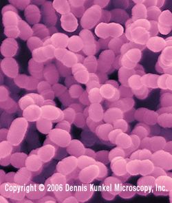
Ticks perch in low vegetation and wait for a susceptible host on which they can attach and feed on. Various online sources describe how ticks enter a host cell. First the R. Rickettsii attach to a protein-dependent receptor on the cell membrane of the host. The cell wall of R.Rickettsii is composed of the outer membrane, peptidoglycan, and cytoplasmic membrane. This makes it hard to stain with the Gram stains and view under the microscope. Secondly, with the aid of the outer membrane Protein A (ompA), the adhesion of this molecule to the host cell induces the local cytoskeleton arrangements with the cell, which results in their entry into the cell. Once a tick attaches to its host, some are known to secrete a cementing material to fasten themselves to the host. Some ticks secrete an anticoagulant, immunosuppressive, and anti inflammatory substance into the area of the tick bite to help the tick obtain a blood meal without the host noticing. The same substances help any freeloading pathogens to establish a foothold in the host.

The Damage following R. Rickettii happens in the blood vessels of the human body, mostly in the brain, skin, and the heart. The bacteria are able to live in the cytoplasm of the nucleus of the host cell. When that bacteria replicate, the results are severe damage and often death of the cells in which it lives in. During multiplication, blood leaks out into nearby tissues through holes in the vessel walls. This obstructs the flow of blood. This part of the cycle involving injury to the blood vessels causes the rash associated with Rocky Mountain spotted fever, in addition to other symptoms including stupor and terminal shock. Death is often caused through excessive damage to the endothelial cells, resulting in the leaking of plasma, decrease in blood volume and shock.

Ticks can transmit many pathogens such as; bacteria, spirochetes, Rickettsia, protozoa, viruses, nematodes and toxins. Tick bite contraindications can resemble arthritis, or flulike symptoms, so it is always a good idea to check yourself anytime after a camping event, or if you have animals that may carry them on their fur. Taking the time to check yourself can prevent a wide variety of symptoms from something as simple as a rash or to something as dangerous as paralysis, shock, or death. Some bites can be cured with and antibiotic, unless it has invaded the Central Nervous System.

References:
1.) http://microbewiki.kenyon.edu/index.php/Rickettsia_rickettsii

2.)http://www.bio.davidson.edu/people/sosarafova/Assets/Bio307/liwoeste/PathogenLifeCycle.html

3.)http://emedicine.medscape.com/article/786652-overview

Sunday, April 17, 2011

Herpes Simplex Virus Type 2

As one of the most common sexually transmitted diseases world wide, Herpes Simplex Virus-2 is without question a public health concern, (approximately 536 million are HSV-2 positive world wide.) Surprisingly though, recent findings reveal that, unlike what most of us think we know about STD's, one does not have to present with symptoms of the virus to be actively contagious in the community.   According to a study presented at a JAMA media research convention, Dr. Anna Wald, (University of Washington and Fred Hutchinson Cancer Research Center), discussed that "the risk of sexual transmission does not correlate with the recognition of clinical signs and symptoms of HSV-2 but most likely correlates with the activity of the virus on the genital skin or mucosa (viral shedding.)"

Data was collected from studies performed by Dr. Anna and her colleagues from 1992 to 2008, of 498 seropositive HSV-2 carriers. Polymerase Chain Reaction was performed on samples from these studies, (collected for 30 consecutive days), of the genital mucous of these individuals for testing of the viral DNA. Subclinical genital shedding rates along with the PCR testing revealed that "...the median [midpoint] amount of HSV detected ...was similar in persons with symptomatic and asymptomatic infection." The same studies also revealed that those individuals presenting lesions (symptoms) were indeed infectious while symptoms lasted (43% of the total amount of days they were tested for, carriers were shedding), but asymptomatic persons only presented 16.4% of the the time (in testing.)  These findings demonstrated that asymptomatic individuals were still at risk for "shedding" or passing on the infection, despite an absence of visible lesions.


Although it is not fully understood how the herpes virus invades cells, it is understood that "most viruses need cell-entry proteins called fusogens in order to invade cells," but herpes requires fusogens plus two other "entry" proteins to invade cells.  Efforts to combat the disease are ongoing, and research and experiments performed by many scientists including those of Dr. Ekaterina Hedwein have "led us to believe that this protein complex is not a fusogen itself but that it regulates the fusogen. We also found that certain antibodies interfere with the ability of this protein complex to bind to the fusogen, evidence that antiviral drugs that target this interaction could prevent viral infection."
As many already know, there is no cure for the herpes virus and it persists in the body for the infected person's lifetime, retreating or resurfacing at random and irregular intervals.  Known effects of the virus range from cold sores, encephalitis, blindness, cancer, and in cases of transmissions from mother to fetus, even death (of the fetus).  Hence, despite what we think we already know about risks of transmission and keys to avoiding infection, ongoing research is imperative to finding a cure for those who, for one reason or another, have contracted the virus

 insciences.org/article.php?article_id=9255

http://www.sciencedaily.com/releases/2011/04/110412101324.htm

lib.jiangnan.edu.cn/ASM/385-2.jpg

Toothbrush Bacteria


Have you ever wondered what it would be like to only have to brush your teeth once a week or even once a month? Well, scientists have identified an enzyme in the human mouth that prevents the buildup of plaque which inevitably leads to tooth decay. The interesting part of this is that this enzyme is naturally produced in our mouths.


The human mouth is full of bacteria that are helpful to our well-being. These bacteria break down the food we eat, keep our mouths clean, and fight of certain infections. The main bacteria that cause plaque, Steptococcus mutans, produce acid from the sugars that we eat. This acid eventually wears down the main protective covering of our teeth known as enamel. Once the enamel is gone, there is no coming back. Conveniently, this is not the only species of bacteria that inhabit our mouths. The good kind, S. salivarius, inhibit the buildup of the bad kind of bacteria, S. mutans.


Through specific tests and careful observations, scientists were able to detect what exactly caused the “good” bacteria to inhibit the spread of the “bad” bacteria. What they found was that a certain enzyme known as, FruA, breaks down sugars thus preventing the buildup of plaque. With these findings, people may believe that they can eat all the candy they wants, but that is not the case. Sucrose, the sugar most commonly ingested by humans, was shown to prevent the “good” bacteria from inhibiting the bad bacteria.


Now, all these scientists have to do is implement this enzyme into toothpastes. The only problem they have is developing a way to keep the enzyme active on the shelf like toothpastes available now. They do believe that the recent information regarding these enzymes will lead to the development of better toothpaste. When they develop these new toothpastes, dentists might start going out of business because no one will need them again!


http://news.sciencemag.org/sciencenow/2011/04/a-bacterium-that-acts-like-a-toothbrush.html


Images:


http://microbewiki.kenyon.edu/images/thumb/5/52/26643C.jpg/250px-26643C.jpg


http://www.orientalcures.com/Images/ToothDecay.jpg


Proteus Vulgaris


Proteus Vulgaris is a Gram negative bacilli shaped bacterium with an extracytoplasmic outer membrane that is believed to cause serious infections in humans. It is a part of the Enterobacteriaceae family as well as Escherichia coli, Klebsiella pneumonia, Enterobacter cloacae, and Serratia marcescens. The Proteus species in general is usually found in the intestinal flora of the intestinal tract, but also in nursing homes, assisted living residences, hospitals, soil, water and plants as well. In hospitals and long care facilities, Proteus finds its way into the patients and employees by the skin and oral mucosa. Proteus mirabilis is the culprit of 90% of all Proteus infections and can be classified as a community acquired infection.


“Infection depends on the interaction between the infecting organism and the host defense mechanisms. Various components of the membrane interplay with the host to determine virulence.” Another important factor in infection is the positive correlation of its size. Actually being infected with a bacterium is a process. The first step of that process it the virus/ bacteria infecting the host cell/ tissue. Once the tissue has been infected, the bacterium has to adhere to it using pili and chemical receptors. Once the bacterium is completely adhered, chemical messengers are sent out as a response, usually causing an infection. Certain chemicals in the body will react with bacteria to form different infections. For example, urease production coupled with bacteria can cause UTI’s (urinary tract infections). Proteus vulgaris is associated with about 35% of all urinary tract infections. “The ability of Proteus organisms to produce urease and to alkalinize the urine by hydrolyzing urea to ammonia makes it effective in producing an environment in which it can survive.”


Proteus vulgaris also has distinct characteristics that set it apart from other Gram negative bacteria. One feature is that they are very motile and literally “swarm” across the plate. This causes a thin film to form on top of the bacteria and a slight color change occurs. There are periods of rest in this “swarm” where they have to slow down and grow and divide. Since they only slow down, and not completely stop, this period of rest is called “swimming”. The other feature that they possess is the ability to turn urea into ammonia using urease. When this reaction occurs, and urease is utilized, the color of the medium changes from red to yellow.


Is Your Meat Contaminated?


Drug resistant strains of Staphylococcus aureus have been found in the meat and poultry of U.S. grocery stores at incredibly high rates. S. aureus is a bacteria known for causing a wide variety of human diseases. Although proper cooking of contaminated meats usually kills the bacteria, there are big issues with cross-contamination.


47% of all meat and poultry sampled were contaminated with S. aureus, and 52% were resistant to three or more antibiotics. Researchers analyzed over 80 brands of meat in 26 grocery stores.


"For the first time, we know how much of our meat and poultry is contaminated with antibiotic-resistant Staph, and is is substantial," said Lance B. Price, Ph.D., senior author of the study and Director of TGen's Center for Food Microbiology and Environmental Health.


On densely-stocked industrial farms, animals are steadily fed low doses of antibiotics make for ideal breeding ground for the bacteria to grow. Even though the U.S. government routinely checks for contamination of our foods, S. aureus is not one of the bacteria they inspect for. S. aureus can cause minor skin infections to life-threatening disease.


"Antibiotics are the most important drugs that we have to treat Staph infections; but when Staph are resistant to three, four, five, or even nine different antibiotics--like we saw in this study--that leaves physicians few options," Dr. Price said.


Sunday, April 10, 2011

Helicobacter pylori: The Cause of Peptic Ulcers

Approximately one-sixth of the world's population suffers from stomach (peptic) ulcers caused by the hard to treat microbe, Helicobacter pylori (H. pylori). A peptic ulcer is a sore on the lining of the stomach or the duodenum. A common misconception of ulcers are that they come from stress or eating spicy foods, but it is H. pylori that cause these painful sores. The bacterium causes peptic ulcers by damaging the mucous coating that protects the stomach and duodenum. When the mucous coating is damaged it then allows stomach acid to get to the sensitive lining beneath. H. pylori is thought to be obtained through food that has not be washed well or cooked properly or from drinking water that has come from an unclean source. It is also thought that an infected person can spread the bacterium to an uninfected person. Although researchers are still unclear how this works it is thought that it can be passed by an uninfected person coming in contact with the stool or vomit of an infected person. It is also thought that it can be passed through the direct contact of saliva.

If a person is thought to have a peptic ulcer caused by H. pylori a doctor can do three noninvasive tests to test for the bacterium. The first is a blood test that checks for H. pylori antibodies. The second is a urea breath test. The patient swallows a capsule, liquid, or pudding that contains urea labeled with a specific carbon atom. After a few minutes the patient breathes into a container, exhaling carbon dioxide. If the carbon atom is found in the breath then H. pylori is present. This is because the bacterium contains a large amount of urease. The third test is a stool antigen test which tests for H. pylori antigens in the patient's stool.

To kill the H. pylori researchers have found that they need to block a key chemical pathway that the bacteria needs for survival. Flabodoxin, a key protein that H. pylori needs for survival, happens to be what needs to be blocked. However, the problem is that H. pylori eaisly becomes resistant to certain antibiotics. Sancho and his team screened 10,000 chemicals for their ability to block flavodoxin and only identified four that showed promise. Three of the four substances killed the bacterium and did not have any apparent toxic effects in lab animals. It is now believed that in order to get rid of H. pylori the antibiotic clarithromycin, a PPI, and the antibiotics amoxicillin or metronidazole for 10 to 14 days will do the trick.


Electric Microbes For Alternative Energy Use


When we think about microbes, we think of tiny organisms that are too small to be seen with the naked eye. Microbes can vary from being deadly to life saving. Microbes are diverse and unique, and we continue to learn more about them everyday. Electric microbes are organisms being looked into as an alternative energy source. These interesting tiny microbes are known as Geobacter. Alternative energy researchers are excited by this discovery. The conductive ability of these tiny particles were discovered by researchers at the University of Massachusetts, and named by Time Magazine as one of the best 50 inventions of 2009.


What are Electric Microbes?

Electric Microbes are colonies of bacteria, that individually communicate and transport energy between one another by sending electrical impulses through cilia, which are tiny hairs growing on the surface of bacteria. The survival of the colony is supported by the electrical connection between the microbes. Just like electrical appliances these microbes function as long as their is an electrical connection traveling through the nanowires. The microbes can share energy simply through touch. In their natural sediment environment the cilia of Geobacter helps it to produce electric current from mud and waste water.


How Electric Microbes are Contributing to Alternative Energy Research?

When colonies of electric microbes exists in envirionments that do not have enough oxygen, they digest food differently. Rather than breaking down sugars to form carbon dioxide and water, the microbes produce carbon dioxide, protons, and electrons. The electrons move through the colony, producing electric current. The current produced is not as strong as the current that runs through household wiring but is significant enough to allow the microbes to survive. Alternative energy researchers are focusing on the capability of electric microbes to alter organic material into electricity. Researchers are creating fuel cells that mock the conversion process these microbes use. But the electricity that is produced by the microbes are transported into an alternative power source rather than for the essential life functions of Geobacter. Microbial fuel cells are unique and versatile, compared to other types of fuel cells. Conventional fuel cells only function using one type of fuel, hydrogen, compared to fuel cells generated using electric microbes, they have the ability to use a wider variety of fuel sources, as long as they are organic and water-based.


Use of Electric Mcirobes Happening Now

These electric microbes are getting more and more attention as new fuel cell ideas are tested. The United Nations plans to test microbial fuel cells in space. As a result of this research, a new power source on the space station would be created by transforming human waste into electrical power. Microbial fuel cells are already being used to amplify some other compelling undertakings, ranging from a self feeding robot to basic fuel cells to help provide lighting in developing countries. These microbes are attractive possibilities for energy sources because they're cheap and easy to maintain. As technology and the understanding of electric microbes advance, more interesting ideas will be developed.


Thursday, April 7, 2011


Food Microbiology

By: Abby VanFossen


Food microbiology is the study of the microorganisms that inhabit, create, or contaminate food. Of major importance is the study of microorganisms causing food spoilage.1 "Good" bacteria, such as probiotics, are becoming increasingly important in food science.2 Also, microorganisms are essential for the production of foods such as cheese, yogurt, other fermented foods, bread, beer and wine.

Food safety is a major focus of food microbiology. Pathogenic bacteria, viruses, and toxins produced by microorganisms are all possible contaminants of food. However, microorganisms and their products can also be used to combat these pathogenic microbes. Probiotic bacteria can kill and inhibit pathogens. Also, bacteriophages, viruses that only infect bacteria, can be used to kill bacterial pathogens. Thorough preparation of food, including proper cooking, eliminates most bacteria and viruses. However, toxins produced by contaminants may be heat resistant, and some are not eliminated by cooking.


Probiotics are living organisms that, when consumed, have beneficial health benefits outside their nutritional effects. There is a growing body of evidence for the role of probiotics in gastrointestinal infections, irritable bowel syndrome and inflammatory bowel disease.3


Lactobacillus species are used for the production of yogurt, cheese, sauerkraut, pickles, beer, wine, cider, kimchi, chocolate and other fermented foods, as well as animal feeds such as silage. In recent years, much interest has been shown in the use of lactobacilli as probiotic organisms and their potential for disease prevention in humans and animals.4


1 Fratamico PM and Bayles DO (editor). (2005). Foodborne Pathogens: Microbiology and Molecular Biology. Caister Academic Press.


2 Tannock GW (editor). (2005). Probiotics and Prebiotics: Scientific Aspects. Caister Academic Press.


3 Ljungh A, Wadstrom T (editors) (2009). Lactobacillus Molecular Biology: From Genomics to Probiotics. Caister Academic Press.


4 Mayo, B; van Sinderen, D (editor) (2010). Bifidobacteria: Genomics and Molecular Aspects. Caister Academic Press.

Genetic Rearrangement in Mice


A study was done in mice to find hotspots of genetic recombination, which is places in the DNA that break and reorganize to create new genes. The ultimate goal of the study was to find out how the recombination of genes affected the rest of the genome of an organism. Their findings are significant in that this may help to show how detecting certain genes can be linked to some diseases or it can also help in detecting abnormalities within the genes.


The scientists used mice in the experiment and created one of the first maps of these genes for a multi-cellular organism. In order to create this map, researchers combined small pieces of DNA they took from the mice that were going through the process of recombination at that time. The map showed that the rearrangement of the chromosomes in the gene has the potential to make each cell identical, which would pose a problem. Using this map, the researchers hope to be able to determine how these abnormalities occur.


The map provided the researchers with green colors, symbolizing the chromosomes, and red colors, symbolizing areas where the chromosomes were most likely going to break apart. These colors displayed to the researchers where variations and abnormalities could occur within the DNA. Even though this study was done in mice, it will be beneficial for humans when determining where certain mutations and abnormalities come from within a gene.


References:


Monday, April 4, 2011

Cryptococcus: A fungus that loves the sugar in your brain

Cryptococcus is a genus of fungus and it grows in a culture in the form of a yeast. Usually a fungus only has about two genes but Cryptococcus has about twelve. This large number of genes allows this fungus to borrow inositol from a persons' brain by encoding for sugar transport molecules. Inositol is a sugar found in the human brain, as well as in the spinal cord. The yeastlike fungi consume inositol, because it's a sugar and because sugar, especially this one, helps it reproduce and more specifically to reproduce sexually. This fungus sexually reproduces because, "A connection between the high concentration of free inositol and fungal infection in the human brain,"which was stated by Chaoyang Xue, Ph.D.This fungus then has a fondness to infect the brain and possibly cause meningitis or other infections. Although before this fungus was interested in the brain, it found inositol on plants in the wild. M.D.,  Ph.D., Joseph Heitman, who is the chairman of the Duke department Molecular Genetics and Microbiology stated that this fungus, "…has the machinery to efficiently move sugar molecules inside of its cells and thrive." Scientists have thought of a way to possibly prevent Cryptococcus infections by putting them on a fungal equivalent of an Atkins diet; known as a low-carb diet. This would deter the sugar loving fungus from multiplying which in return would possibly stop infections.


Cryptococcus.jpg


http://www.sciencedaily.com/releases/2010/04/100405152757.htm

http://www.sott.net/articles/show/206336-This-Is-Your-Brain-on-Cryptococcus-Pathogenic-Fungus-Loves-Your-Brain-Sugar

http://www.google.com/search?hl=en&defl=en&q=define:Cryptococcus&sa=X&ei=y22aTZriDeLX0QHh4YTHBg&ved=0CBQQkAE